About Retina
The retina converts light that enters into your eye into electrical signals your optic nerve sends to your brain which creates the images you see. It’s a key part of your vision. The retina is the layer at the very back of your eyeball.
Examining the retina
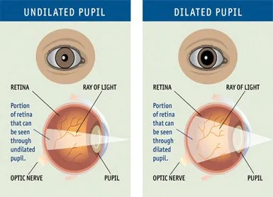
A retinal examination also called as ophthalmoscopy or funduscopy. It allows the retinal specialist to evaluate the back of your eye, including your retina, optic disk and the underlying layer of blood vessels that nourish the retina (choroid).
For a through retinal examination the pupils must be dilated using eye drops. The eyedrops used for dilation cause your pupils to widen, allowing in more light and giving your doctor a better view of the back of your eye.


Who should get a dilated retinal examination?
The following are some of the indications for frequent dilated eye examination:
- Diabetes
- High blood pressure
- Recent onset of sudden of progressive vision loss
- Family history of retinal detachment
- High spectacle powers
- Recent onset of flashes or floaters in the vision.
- Distortion in size and shape of objects.
- Everyone at the age of 40 years
- On drugs for Rheumatic problems.
- Injury to the eye
How is it treated?
Huge strides have been made in the treatment of diabetic retinopathy. Treatments such as scatter photocoagulation, focal photocoagulation, and vitrectomy prevent blindness in most people. The sooner retinopathy is diagnosed, the more likely these treatments will be successful. The best results occur when sight is still normal.
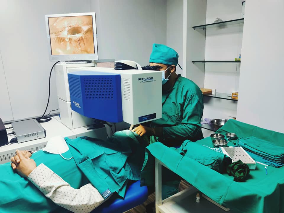
- In photocoagulation, the eye care professional makes tiny burns on the retina with a special laser. These burns seal the blood vessels and stop them from growing and leaking.
- In scatter photocoagulation (also called panretinal photocoagulation), the eye care professional makes hundreds of burns in a polka-dot pattern on two or more occasions. Scatter photocoagulation reduces the risk of blindness from vitreous hemorrhage or detachment of the retina, but it only works before bleeding or detachment has progressed very far. This treatment is also used for some kinds of glaucoma.
- Side effects of scatter photocoagulation are usually minor. They include several days of blurred vision after each treatment and possible loss of side (peripheral) vision.
